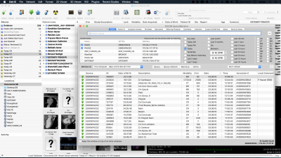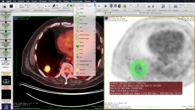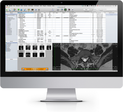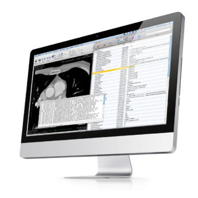
#Features osirix md software
We will compare the features of the VITREA2® and AW VolumeShare 5® radiology workstation with free open source software applications like OsiriX® and 3D Slicer®, with examples from specific studies. With this study, we aim to present the advantages and disadvantages of these radiological image visualization systems in the advanced management of radiological studies. Nevertheless, the programs included in these workstations have a high cost which always depends on the software provider and is always subject to its norms and requirements. These radiological devices are basically CT (Computerised Tomography), MRI (Magnetic Resonance Imaging), PET (Positron Emission Tomography), etc. In radiology, graphic workstations allow their users to process, review, analyse, communicate and exchange multidimensional digital images acquired with different image-capturing radiological devices. Consequently, sophisticated computing tools that combine software and hardware to process medical images are needed. For the radiological field, manipulating and post-processing images is increasingly important.

However, this last group of free applications have limitations in its use. Two examples of free open source software are Osirix Lite® and 3D Slicer®.
#Features osirix md code
In addition, some of these applications are known as Free Open Source Software because they are free and the source code is freely available, and therefore it can be easily obtained even on personal computers. These software applications are of great interest, both from a teaching and a radiological perspective.

Conclusion The new software is a promising tool for coronary CTA post-processing providing good overviews of the coronary artery with limited user interaction on low-end hardware, and the coronary CTA diagnosis procedure could potentially be more time-efficient than using thin-slab technique.Currently, there are sophisticated applications that make it possible to visualize medical images and even to manipulate them. Visually correct centerlines were obtained automatically in 94.7% (321/339) of the intact branches. Results The median processing time was 6.4 min, and 100% of main branches (right coronary artery, left circumflex artery and left anterior descending artery) and 86.9% (219/252) of visible minor branches were intact. The software was evaluated in 42 clinical coronary CTA datasets acquired with 64-slice CT using isotropic voxels of 0.3–0.5 mm. The main segmentation function is based an optimized “virtual contrast injection” algorithm, which uses fuzzy connectedness of the vessel lumen to separate the contrast-filled structures from each other. Materials and Methods The software was built as a plug-in for the open-source PACS workstation OsiriX. Object A new software module for coronary artery segmentation and visualization in CT angiography (CTA) datasets is presented, which aims to interactively segment coronary arteries and visualize them in 3D with maximum intensity projection (MIP) and volume rendering (VRT). This allows conservators to access different forms of information and to view a variety of image types simultaneously.

The software we present is designed and structured to accommodate multiple types of digital imaging data, including as yet unspecified or unimplemented formats, in an integrated environment. Second, previous software has largely allowed visualizing a single type or only a few types of data. We provide an open-source software tool with a single intuitive interface consistent with conservators' needs that handles various types of 2D and 3D image data and preserves user-generated metadata and annotations.

First, users must deal with a wide variety of interfaces specialized for applications besides conservation. In this paper, we address two practical barriers to using high-tech digital data in art conservation. In addition, many of these software packages are focused on particular applications (such as medicine or remote sensing) and do not permit users to access and fully exploit all the information contained in the data.
#Features osirix md professional
However, viewing most of this digital image data frequently requires both specialized software, which is often associated with a particular type of acquisition device, and professional knowledge of and experience with each type of data. Commonly used techniques include hyperspectral imaging, 3D scanning, and medical computed tomography imaging. Art conservators now have access to a wide variety of digital imaging techniques to assist in examining and documenting physical works of art.


 0 kommentar(er)
